Documented digital health technology · 04
TrichoLens: A FolliGenz Technology for Hair and Scalp Screening
How TrichoLens combines calibrated image capture, computer vision, structured hair metrics, manual measurement, progress photography, and AI-assisted explanations to help users observe change over time without presenting the platform as a diagnostic service.
- Organizations
- FolliGenz Therapeutics Corporation and TrichoLens
- Project period
- March 2026 methodology, platform reviewed July 2026
- Prepared by
- Biotica Consulting LLC
- Last reviewed
- July 31, 2026
- Status
- Live screening technology
- Format
- Image-based screening and longitudinal tracking platform
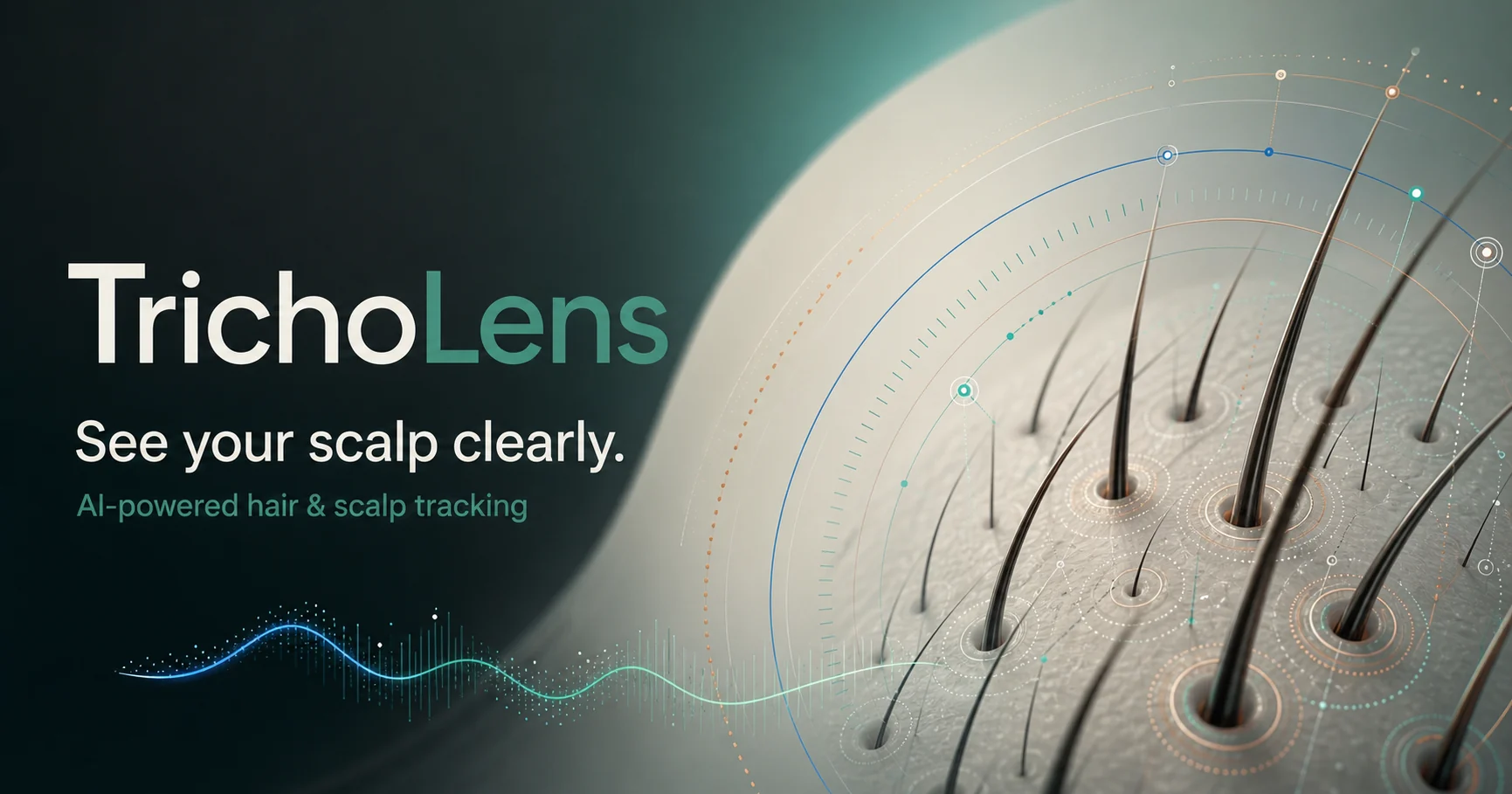
TrichoLens is presented here as a FolliGenz technology. The supplied March 2026 methodology uses the earlier working name Dermolens throughout, while the current platform and official social-share image use TrichoLens. Biotica Consulting prepared this technical case-study summary. The methodology does not identify a software developer, validation laboratory, or medical reviewer, so this page does not assign those roles or claim clinical validation.
A screening system designed around repeatable observation
Hair and scalp changes are difficult to judge from memory or from photographs captured under different conditions. TrichoLens addresses that practical problem by turning trichoscopy images, close-up scalp photographs, and repeat progress photos into a structured record. The platform is designed to identify visible hairs, estimate width and density, organize image-derived metrics, and help users compare the same areas across time.
The methodology describes a layered workflow. Users first provide an image and, when possible, the physical field of view. A computer-vision model detects visible hairs and produces segmentation polygons. Geometry from those polygons is converted into estimated hair width, then grouped into methodology-defined terminal, intermediate, and vellus bands. The system derives screening indicators such as estimated density, terminal-to-vellus ratio, Hair Maturity Index, and a composite Hair Count Rating. Manual measurement, spot analysis, self-assessment, scalp scanning, image galleries, and a guided Photo Diary extend the record beyond a single automated scan.
The strongest use case is longitudinal screening with consistent capture conditions, not one-time diagnosis. Results depend on lighting, focus, magnification, field-of-view calibration, device characteristics, hair color contrast, and model performance. The methodology is a technical description, not an independent validation report. It contains no clinician-ground-truth study, diagnostic accuracy analysis, external validation, or regulated medical-device claim. TrichoLens should therefore be described as an awareness and tracking tool that can help users prepare better questions for a qualified professional.
Clear ownership and careful attribution
FolliGenz Therapeutics Corporation
TrichoLens is identified for this case study as a FolliGenz technology, extending the company's hair and scalp research interests into structured digital screening and progress tracking.
TrichoLens
Provides image capture, computer-vision estimates, measurement tools, AI-assisted explanations, longitudinal photo workflows, and user-facing records. The platform states that it is for screening and does not replace professional medical evaluation.
Biotica Consulting LLC
Reviewed the supplied methodology, separated documented functions from unproven clinical claims, and translated the technical workflow into this public evidence-bounded case study.
From an image to a repeatable progress record
- 01
Start with a defined field of view
Users upload trichoscopy, close-up scalp, or standard phone images. Known physical dimensions improve scaling; otherwise, the system uses a documented default assumption and warns that device and magnification differences can affect the estimate.
- 02
Convert visible image structure into screening metrics
Detection, segmentation, geometric width estimation, classification, and plausibility checks produce structured indicators. Manual tools let users review selected shafts and visible scalp regions directly.
- 03
Repeat capture under comparable conditions
Photo Diary guides, prior-image overlays, lighting checks, multi-zone views, saved reports, and timeline media are intended to make relative change easier to observe over weeks and months.
Questions the work was designed to answer
- How can a consumer image be translated into structured hair and scalp screening estimates without presenting those estimates as a diagnosis?
- Which calibration assumptions determine the credibility of width and density outputs?
- How does the computer-vision pipeline identify, segment, classify, and count visible hairs?
- Which fallback metrics remain useful when a terminal-to-vellus ratio is unstable or unavailable?
- How can guided photography reduce noise when users compare images over time?
- What performance, privacy, and clinical evidence is still needed before stronger claims are appropriate?
What the documented technology is designed to do
Calibration is the foundation of every physical estimate
Users can enter the physical width and height represented by an image. When those values are unknown, the methodology assumes a 2.5 mm by 2.5 mm field for a square image and scales non-square images proportionally. This supports an order-of-magnitude estimate, not a device-independent measurement. Width and density become more credible when the actual field of view is known.
Computer vision prioritizes visible thin hairs
The documented detector returns bounding boxes and segmentation polygons for visible hairs in trichoscopy images. It is tuned to favor sensitivity so finer hairs are less likely to be omitted, and overlap filtering reduces duplicate counts. Lighting, focus, contrast, compression, overlapping shafts, and very lightly pigmented hair can still change detection performance.
PCA converts segmentation geometry into estimated shaft width
For each detected polygon, Principal Component Analysis identifies the major axis associated with hair direction. Polygon points are projected onto the perpendicular axis, and that span becomes the width in pixels. The selected calibration then converts pixels into a physical estimate. This is image-derived geometry, not controlled laboratory microscopy.
Classification bands organize possible miniaturization signals
The methodology groups estimated widths into terminal at 60 micrometers or greater, intermediate from 30 to under 60 micrometers, and vellus below 30 micrometers. It also describes user-selected reference bands for Asian, Caucasian, and African hair. Because the document provides no bibliography or validation dataset for those values, they should be described as methodology-defined screening bands, not universal clinical cutoffs.
Plausibility checks look for likely scaling errors
Measurements below a documented visibility floor or above a selected reference maximum trigger calibration guidance. In magnification mode, the system can suggest an adjustment while prioritizing the thinnest measured hair. In manual field-of-view mode, it asks the user to verify dimensions. These checks catch implausible outputs, but they do not replace calibration against a known physical reference.
Multiple metrics prevent one ratio from carrying the whole interpretation
The terminal-to-vellus ratio compares classified terminal and vellus hairs. When fewer than three terminal hairs make that ratio unstable, the methodology uses a Hair Maturity Index that weights terminal hairs at 1.0, intermediate hairs at 0.5, and vellus hairs at 0.0. An intermediate-to-vellus ratio provides another view of the classified distribution. These are screening summaries, not validated disease-severity scores.
Density and the composite rating inherit calibration uncertainty
Estimated density divides detected hairs by the image area derived from the selected scale. The Hair Count Rating then combines 60 percent normalized density and 40 percent Hair Maturity Index. A clean formula makes the output understandable, but an incorrect field of view or missed detections will propagate into both measures.
Manual and focused tools keep users inside the evidence loop
The Hair Thickness tool lets users place measurement points across selected shafts, edit anchors, group measurements into segments, and export annotated images. Spot Analysis uses brightness thresholds and connected-component labeling to mark visible regions, while Self Assessment records user-rated symptoms. A detected spot does not establish its biological cause, and a self-rating is not a clinical examination.
Photo quality and repeatability are treated as measurable inputs
Standard photos can be checked for brightness and sharpness before AI review. The Photo Diary supports front, left, right, and top views, framing silhouettes, a low-opacity overlay of the previous image, and lighting comparisons against prior captures. These controls are important because repeatable photography may be more informative than a visually impressive but inconsistent one-time image.
AI explains structured outputs but does not convert them into a diagnosis
The methodology describes server-side AI features that receive tool-specific images and metrics, generate summaries, and support follow-up questions. Server-side credential handling protects an API key, but it does not by itself prove privacy compliance, clinical accuracy, or safe handling across every jurisdiction. AI observations remain dependent on the image and the upstream measurements.
The platform is strongest as a longitudinal screening record
The methodology repeatedly emphasizes consistent device, magnification, lighting, angle, and field of view. Under those conditions, trends within the same user's record may be more meaningful than a single absolute estimate. The document does not provide precision, recall, clinician agreement, device reproducibility, or prospective outcome data.
Evidence needed before clinical or performance claims
The project record is a development input, not permission to outrun the evidence. These are the next questions for direct testing and review.
- How does the detector perform on an independent test set across skin tones, hair colors, curl patterns, scalp zones, devices, and image-quality conditions?
- What are precision, recall, segmentation quality, duplicate rate, and missed-hair rate against expert-annotated ground truth?
- How closely do width and density estimates agree with calibrated microscopy or a validated phototrichogram method?
- What are repeatability and reproducibility when the same user, a different user, or a different device captures the same region?
- Have the methodology-defined classification bands, fallback ratios, and composite rating been validated against independent clinical assessments?
- How are confidence, uncertainty, low-quality images, insufficient hair counts, and model failure communicated to users?
- What human review and escalation controls address outputs that could be interpreted as diagnostic or urgent?
- What consent, retention, deletion, encryption, access-control, incident-response, and cross-border data rules apply to health-adjacent images and conversations?
- Which intended-use and jurisdictional analyses support the platform's current screening positioning and future professional or enterprise use?
- Can a prospective longitudinal study show that standardized capture improves trend detection and professional conversations without encouraging self-diagnosis?
Progress stated at the level the work supports
TrichoLens presents a thoughtful technical response to a real measurement problem: hair changes slowly, while casual photos introduce enough variation to hide or imitate progress. Its value is the combination of structured image analysis, transparent screening metrics, manual review tools, and repeatable longitudinal capture. The current methodology supports describing a sophisticated tracking and awareness platform. It does not yet support diagnostic accuracy, laboratory-grade measurement, treatment-response prediction, or replacement of a dermatologist.

FolliGenz connected the screening methodology to a buildable software workflow
Biotica supported the methodology, evidence boundaries, product logic, market questions, and validation roadmap behind the TrichoLens screening and tracking platform.
Project records and public sources
The supplied March 2026 methodology is the primary technical record. The live TrichoLens website provides current product context. Numerical thresholds and internal scores are reported as methodology-defined values because the document does not include a bibliography or an independent validation report.
- 01Private project record
Tricholens Screening Tool Methodology
Supplied FolliGenz project record · Technical methodology, 7 pages, March 2026
- 02View source
TrichoLens official platform
TrichoLens · Official technology website
- 03View source
TrichoLens public methodology
TrichoLens · Official methodology page
- 04View source
TrichoLens official social-share image
TrichoLens · Official Open Graph and social preview asset
- 05View source
About FolliGenz Therapeutics
FolliGenz Therapeutics Corporation · Official company page
Project evidence and literature must be read within their stated methods and limitations. This page is not medical, clinical, regulatory, or legal advice.
Related projects and research
Follow adjacent formulation, partner, supplier, and research questions while keeping each evidence type clearly labeled.

Need a screening workflow turned into a testable platform?
Biotica can connect product research, software engineering, market analysis, evidence boundaries, and a validation roadmap for health-adjacent technology in the United States, Canada, Brazil, China, India, and additional markets.
Discuss a software project